Elena sivan-loukianova

In Johnston’s organ (JO), the antennal chordotonal organ (ch) subserving hearing, we have recently shown that Cut is required for correct ch differentiation as cut null antennal clones show disrupted JO morphology and complete loss of auditory response (Ebacher et al., 2007).
Using the viable cut6 allele, we now show female-biased deafness as a result of morphological defects in the shape of the a2/a3 joint, together with disorganization and reduced numbers of scolopidia (ch sensory units). During development, cut6 mutants show loss of some scolopidia at 24 hrs after puparium formation (APF), but the apical ends of the remaining dendritic caps appear to protrude through the epithelial layer into the space between a2/a3 segments as in control flies. We are using a variety of markers to better define these developmental consequences.
In parallel, we are using UAS-ct-RNAi lines to further examine Cut function in JO. Using the ato-Gal4 driver, we observe a strong effect on hearing by electrophysiological analysis. Loss of cut function reduces the number of JO scolopidia, and affects development and attachment of scolopidia, and alters the morphology of the a2/a3 joint.
To explore possible downstream targets of Cut, we conducted microarray expression analysis of eye-antennal discs to compare gene expression in cut6 vs control flies, dissected at late third instar and either analyzed immediately, or after disc culture for 24 hrs. The expression of the selected genes that were upregulated or downregulated in cut6 was verified by using Real-time PCR.

Education:
Ph.D. - Dept. of Histology, State
Universtiy, St. Petersburg, Russia
current position:
Assistant Research Scientist
Contact:
More Images of my work
The role of Cut in Johnston’s organ development and function

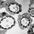
Elena conducts independent research on the ultrastructure of the auditory circuit in normal and mutant Drosophila. The analyses usually include immunohistochemistry at the EM level and the light microscopy (fluorescence, DIC, confocal) level. Elena works on the developmental and cellular mechanisms by dynamic imaging with appropriate markers in mutant and wild -type Drosophila, together with ultrastructural characterization of mutant defects. Also, she uses the culture system of Drosophila eye-antennal discs to investigate control of the most important events in Johnston’s organ development (confocal imaging). Finally, Elena is involved in microarray analysis of early gene expression in antennal discs. Selected genes are later verified by quantitative PCR, in situ hybridization and antibody staining.
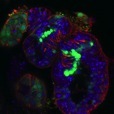
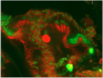
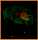
During development, cut6 mutants (right) have fewer scolopidia at 24 hrs APF, but the apical ends of the remaining dendritic caps appear to protrude through the epithelial layer into the space between the a2/a3 segments as in control flies (left).
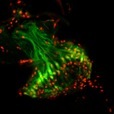
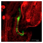
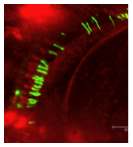
Genotype: wct;ato-Gal4;dicer/ctRNAi
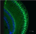
Left: 24 hr APF; Right: adult
Anti-cut –Alexa488 - nuclei
Texas Red Phalloidin – Scolopale rods